 Encyclopædia Neurochirurgica
 Brain arteriovenous malformation


2.3. SM Grades 4 (Figure 10) and 5 (Figure 11) or SP Class C:
Usually conservative treatment. Multimodality treatment is proposed in specific cases such as very young patients, with recurrent hemorrhages or progressive neurological deficit. Partial treatment is also possible in cases of elevated risk factors such as intra-nidal aneurysm and high flow arteriovenous fistulas with progressive neurological deficit. High post-treatment morbidity (deficit risk) rate: 12 to 38%. [25, 32, 95]
The Lawton et al. [47] supplementary grading scale may help in treatment decision. As they proposed, an AVM with a low Spetzler-Martin grade (grade I – III) may be favorable for microsurgical resection, and a low supplementary grade (I – III) may strengthen the recommendation for surgery. Conversely, an AVM with a high Spetzler-Martin grade (IV – V) may be unfavorable for microsurgical resection, and a high supplementary grade (IV – V) may strengthen the recommendation for non-operative management. In cases of matched Spetzler-Martin and supplementary grades, the supplementary grading system has a confirmatory role and may not alter management decisions. However, in cases of mismatched Spetzler-Martin and supplementary grades, the supplementary grading system may alter management decisions and therefore has a more important role.
- Figure 10a
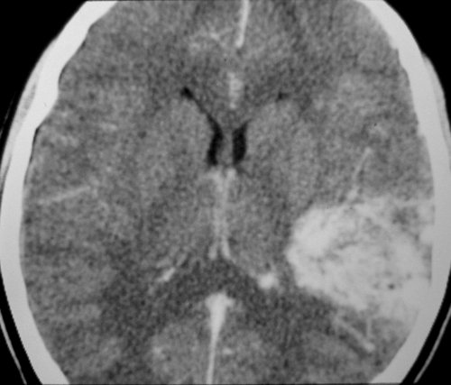
- Grade 4 AVM. a) Axial cerebral CT showing left temporoparietal intraparenchymal hemorrhage secondary to an AVM bleeding.


- Figure 10b
- Grade 4 AVM. b) Angiography in an AP projection of the left internal carotid artery showing an AVM fed by the left ACA and MCA.

- Figure 10c
- Grade 4 AVM. c) Angiography in an AP projection of the left internal carotid artery showing an AVM fed by the left ACA and MCA.

- Figure 10d
- Grade 4 AVM. d) Angiography in an AP projection of the left vertebral artery showing an AVM fed by the left PCA.


- Figure 10e
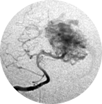
- Grade 4 AVM. e) Angiography in a lateral projection showing an AVM fed by the left MCA.

- Figure 10f
- Grade 4 AVM. f) Postoperative angiography of the left internal carotid artery in an AP projection showing complete ressection of the AVM.


- Figure 10g
- Grade 4 AVM. g) Postoperative angiography of the left internal carotid artery in a lateral projection showing complete ressection of the AVM.

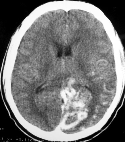
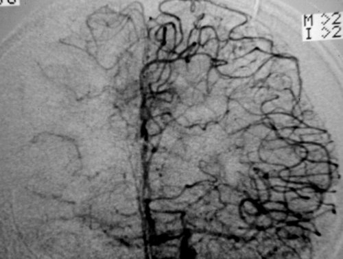
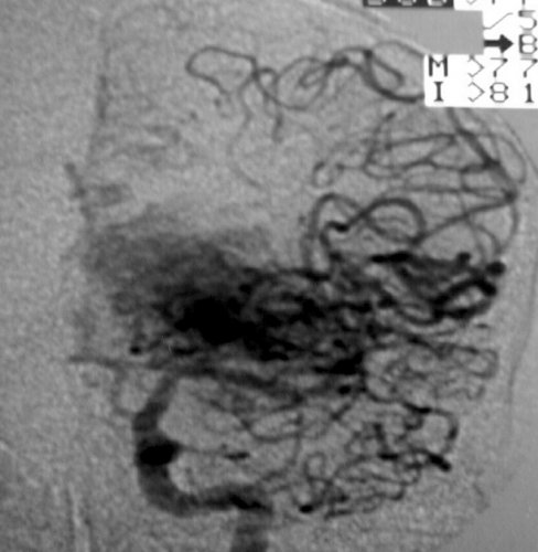
- Figure 11a
- Grade 5 AVM. a) Axial cerebral CT with contrast showing left occipital high density corresponding to an AVM.

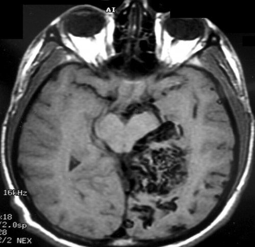
- Figure 11b
- Grade 5 AVM. b) Axial cerebral T1-weighted MRI showing flow void corresponding to an left occipital and mesial temporal AVM.


- Figure 11c
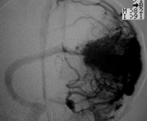
- Grade 5 AVM. c) Angiography in a lateral projection of the left vertebral artery showing an AVM fed by the left PCA.

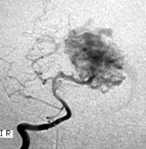
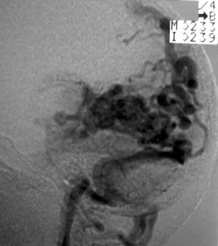
- Figure 11d
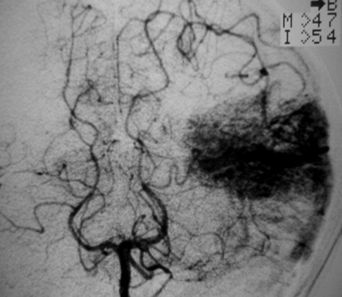
- Grade 5 AVM. d) Angiography in an AP projection of the left vertebral artery showing an AVM fed by the left PCA.

- Figure 11e
- Grade 5 AVM. e) Angiography in an AP projection of the left internal carotid artery showing an AVM fed by the left ACA and MCA.

- Figure 11f
- Grade 5 AVM. f) Late venous phase angiography in an AP projection of the left internal carotid artery showing an AVM with superficial and profound drainage.

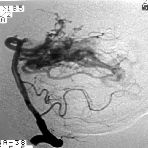
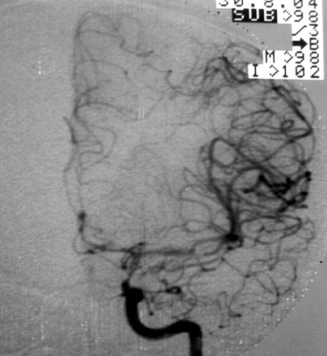
- Figure 11g 1ss emb
- Grade 5 AVM. g) Angiography in a lateral projection of the left vertebral artery after the first embolization section.

- Figure 11h 2ss emb
- Grade 5 AVM. h) Angiography in a lateral projection of the left vertebral artery after the second embolization section.

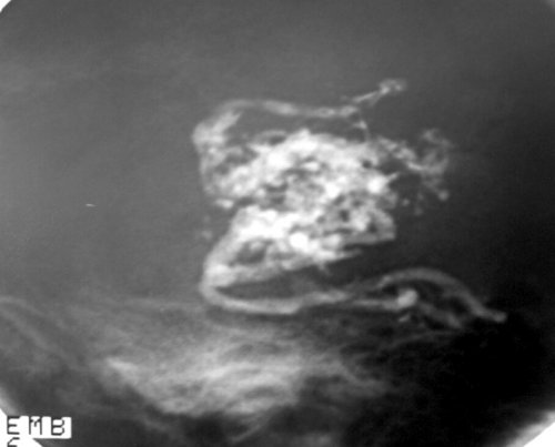
- Figure 11i material emb
- Grade 5 AVM. i) Lateral skull fluoroscopy showing the final embolization cast.


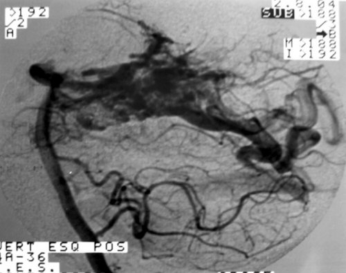
- Figure 11j
- Grade 5 AVM. j) Postoperative angiography of the left vertebral artery in a lateral projection showing complete ressection of the AVM.


- Figure 11k
- Grade 5 AVM. k) Postoperative angiography of the right vertebral artery in an AP projection showing complete ressection of the AVM.

- Figure 11l
- Grade 5 AVM. l) Postoperative angiography of the left internal carotid artery in an AP projection showing complete ressection of the AVM.
