 Encyclopædia Neurochirurgica
 Brain arteriovenous malformation


II. History
Colby et al. [13] performed in 2012 a review of the history of AVMs, from their very beginning until today. We highlight chronologically some of the most fundamental aspects of this long history [13]:![]() Mid-1700s - John Hunter (1728–1793) described the clinical characteristics of extracranial AVMs.
Mid-1700s - John Hunter (1728–1793) described the clinical characteristics of extracranial AVMs.![]() 1863 - Rudolf Virchow (1821–1902) described many of the common intracranial vascular pathologic entities, including AVMs.
1863 - Rudolf Virchow (1821–1902) described many of the common intracranial vascular pathologic entities, including AVMs. ![]() 1889 - Davide Giordano (1864–1954) - the first report of a palliative treatment of a true cerebral AVM by ligation of a left parietal feeding artery.
1889 - Davide Giordano (1864–1954) - the first report of a palliative treatment of a true cerebral AVM by ligation of a left parietal feeding artery.![]() 1889 – Jules-Émile Péan (1830 – 1898) - the first complete excision of a cerebral AVM was made by the famous French surgeon J-E Péan. [112]
1889 – Jules-Émile Péan (1830 – 1898) - the first complete excision of a cerebral AVM was made by the famous French surgeon J-E Péan. [112]![]() 1914 - Vilhelm Magnus (1871–1929) was probably the first to treat cerebral AVMs with radiation using conventional fractionated radiation.
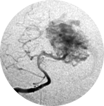
1914 - Vilhelm Magnus (1871–1929) was probably the first to treat cerebral AVMs with radiation using conventional fractionated radiation.![]() 1927 - Antonio Caetano de Abreu Freire Egas Moniz (1874–1955) performed the first successful cerebral angiogram by injecting contrast into the carotid artery of patients. A major advance for the diagnosis and understanding of AVMs.
1927 - Antonio Caetano de Abreu Freire Egas Moniz (1874–1955) performed the first successful cerebral angiogram by injecting contrast into the carotid artery of patients. A major advance for the diagnosis and understanding of AVMs.![]() 1928 - Walter Dandy (1886–1946) and Harvey Cushing (1869–1939) with Percival Bailey (1892–1973) - independently reported a series of AVMs treated before the introduction of angiography, with primarily catastrophic results in both series.
1928 - Walter Dandy (1886–1946) and Harvey Cushing (1869–1939) with Percival Bailey (1892–1973) - independently reported a series of AVMs treated before the introduction of angiography, with primarily catastrophic results in both series.![]() 1932 - Herbert Olivecrona (1891–1980) was the second to successfully remove an AVM. He introduced the technique of ligating superficial feeding vessels and then working in a circumferential fashion until the deep portion of the AVM was dissected and separated from the brain. The AVM draining veins were ligated as a final step.
1932 - Herbert Olivecrona (1891–1980) was the second to successfully remove an AVM. He introduced the technique of ligating superficial feeding vessels and then working in a circumferential fashion until the deep portion of the AVM was dissected and separated from the brain. The AVM draining veins were ligated as a final step.![]() 1932 to 1957 - Gazi Yasargil (born 1925) compiled information from the literature on 500 AVMs: operative mortality for “small” AVMs was 5% and for “moderate-size” AVMs was 10%.
1932 to 1957 - Gazi Yasargil (born 1925) compiled information from the literature on 500 AVMs: operative mortality for “small” AVMs was 5% and for “moderate-size” AVMs was 10%.![]() 1950s - Leonard Malis (1919–1995) devised and constructed bipolar coagulation forceps.
1950s - Leonard Malis (1919–1995) devised and constructed bipolar coagulation forceps.![]() 1951 - Lars Leksell (1907–1986) described the radiosurgery, using a cyclotron.
1951 - Lars Leksell (1907–1986) described the radiosurgery, using a cyclotron. ![]() Early-1960s - Luessenhop and William Spence (1908–1992) - developed largely, endovascular techniques conceptualizing the blockage of hypertrophied, abnormal feeding vessels, by direct puncture of target vessels to introduce catheters and embolic material.
Early-1960s - Luessenhop and William Spence (1908–1992) - developed largely, endovascular techniques conceptualizing the blockage of hypertrophied, abnormal feeding vessels, by direct puncture of target vessels to introduce catheters and embolic material.![]() 1960s - The introduction of the operative microscope and the resulting development of microsurgical instruments and microsurgical techniques
1960s - The introduction of the operative microscope and the resulting development of microsurgical instruments and microsurgical techniques![]() 1966 - George Perret and Hiro Nishioka – initiated the study of AVM natural history reporting an analysis of 545 cases of cerebral AVMs and fistulae with a hemorrhage rate of 1.5%per year.
1966 - George Perret and Hiro Nishioka – initiated the study of AVM natural history reporting an analysis of 545 cases of cerebral AVMs and fistulae with a hemorrhage rate of 1.5%per year.![]() 1968 - Leksell introduced the first Gamma Knife using cobalt sources.
1968 - Leksell introduced the first Gamma Knife using cobalt sources.![]() 1969 - Yasargil – published the first microsurgical AVM series, and his results were excellent.
1969 - Yasargil – published the first microsurgical AVM series, and his results were excellent.![]() 1971 - Atkinson Morley’s Hospital in England – first clinical CT scan on a patient.
1971 - Atkinson Morley’s Hospital in England – first clinical CT scan on a patient.![]() 1974 – Fedor Serbinenko (1928–2002) – in the endovascular field, described the balloon catheterization and occlusion of intracranial vessels.
1974 – Fedor Serbinenko (1928–2002) – in the endovascular field, described the balloon catheterization and occlusion of intracranial vessels.![]() 1976 - Charles Kerber - in the endovascular field, described a calibrated leak balloon system for flow-directed catheter guidance and administration of contrast and cyanoacrylate embolic material.
1976 - Charles Kerber - in the endovascular field, described a calibrated leak balloon system for flow-directed catheter guidance and administration of contrast and cyanoacrylate embolic material.![]() 1977 - Downstate Medical Center in New York - first clinical MRI on a patient.
1977 - Downstate Medical Center in New York - first clinical MRI on a patient. ![]() 1986 - Robert Spetzler and Neil Martin developed the important AVM classification scheme based on lesion size, venous drainage, and eloquence of involved brain.
1986 - Robert Spetzler and Neil Martin developed the important AVM classification scheme based on lesion size, venous drainage, and eloquence of involved brain. ![]() 1990s - development of functional MRI and diffusion tensor imaging to map eloquent areas of brain and their relationship to the lesion.
1990s - development of functional MRI and diffusion tensor imaging to map eloquent areas of brain and their relationship to the lesion.![]() 1991 - Osvaldo Betti and colleagues introduced the radiosurgery using LINAC (linear accelerator)
1991 - Osvaldo Betti and colleagues introduced the radiosurgery using LINAC (linear accelerator)![]() End-2010s - Intraoperative fluorescence video angiography using indocyanine green (ICG).
End-2010s - Intraoperative fluorescence video angiography using indocyanine green (ICG).