 Encyclopædia Neurochirurgica
 Brain arteriovenous malformation


2.2. SM Grade 3 (Figure 9) or SP Class B:
Depending on the size and location, it can be treated with surgery, radiosurgery, embolization, or a combination of these methods (multimodality treatment). The postoperative morbidity (deficit risk) varies from 8 to 16%. [32, 45, 95]
Using the Lawton sub-classification. [45]:
2.2.1. S1V1E1 (III -): surgical risk similar to that of low-grade AVMs (SP A) (new deficit or death: 2.9%) - can be safely treated with microsurgical resection. These lesions may also be treated with radiosurgery. Although, we agree with Lawton et al: when there is a surgical corridor (like the sylvian fissure, interhemispheric fissure, basal cistern, etc) we recommend microsurgical resection. Microsurgical and radiosurgical morbidity rates are often comparable but microsurgery has immediacy and an efficacy of nearly 100%.
Radiosurgery also has high rates of obliteration in small AVM (from 81 to 90%). The main disadvantage of radiosurgery is that complete obliteration takes from 1 to 3 years after the procedure.
2.2.2. S2V1E0 (III): intermediate surgical risks (new deficit or death: 7.1%) - carefully individualized treatment.
2.2.3. S2V0E1 (III +): surgical risk similar to that of high-grade AVMs (SP C) (new deficit or death: 14.8%) - best managed conservatively
2.3.4. S3V0E0 (III*): extremely rare or does not exist
- Figure 9a
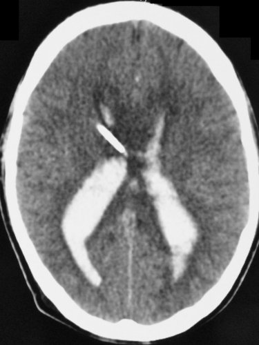
- Grade 3 AVM. a) Axial cerebral CT showing extensive intraventricular hemorrhage secondary to an AVM bleeding and an external ventricular drain catheter in the right lateral ventricle.

- Figure 9b
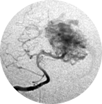
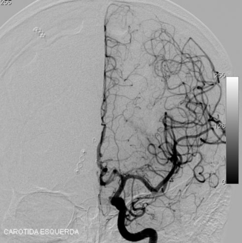
- Grade 3 AVM. b) Angiography in an AP projection showing an AVM fed by a branch of the left ACA and draining to the superior sagittal sinus.

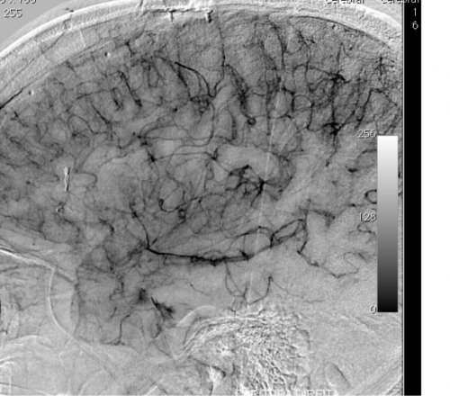
- Figure 9c
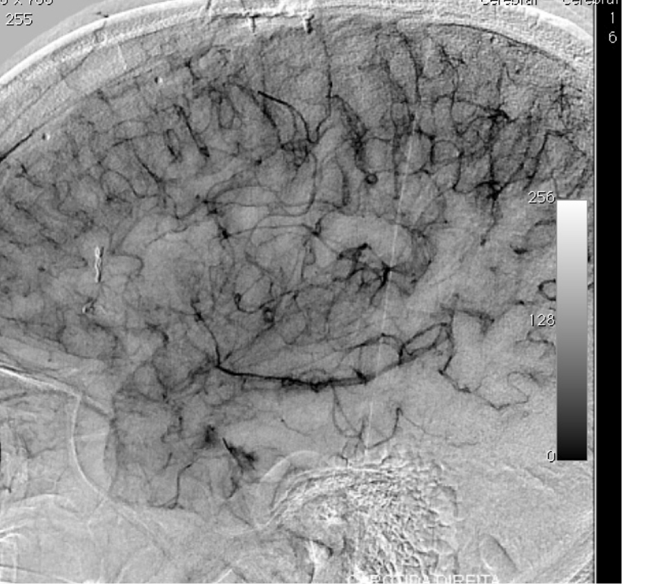
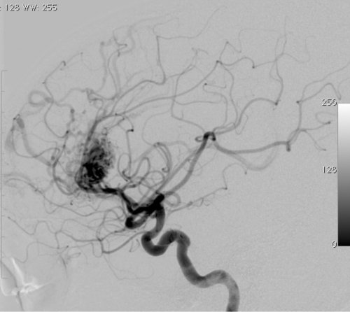
- Grade 3 AVM. c) Angiography in a lateral projection showing an AVM fed by a branch of the left ACA.

- Figure 9d
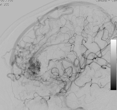
- Grade 3 AVM. d) Late venous phase angiography in a lateral projection showing an AVM draining to the superior sagittal sinus and internal cerebral vein.

- Figure 9e
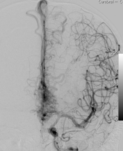
- Grade 3 AVM. e) Postoperative angiography of the left internal carotid artery in an AP projection showing complete ressection of the AVM.


- Figure 9f
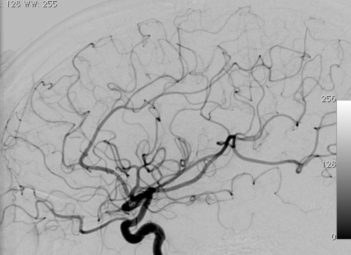
- Grade 3 AVM. f) Postoperative angiography of the left internal carotid artery in a lateral projection showing complete ressection of the AVM.
- Figure 9g
- Grade 3 AVM. g) Postoperative late venous phase angiography of the left internal carotid artery in a lateral projection showing complete ressection of the AVM.