 Encyclopædia Neurochirurgica
 Brain arteriovenous malformation
, popularity : 11%
[English]
[français]


*2. Classification and treatment
The Spetzler-Martin (SM) and Spetzler-Ponce (SP) grading systems [95, 96], as previously described, can be a guide for the therapeutic decision:
2.1. SM Grades 1 (Figure 7) and 2 (Figure 8) or SP Class A:
The treatment of choice is microsurgical resection, but embolization or radiosurgery may be considered as well. In the literature, the risk of morbidity (deficit risk) in the postoperative period ranges from 0.7 to 5% for microsurgery. [25, 32, 92, 95] Whereas preoperative embolization in class A is associated with a fall in the modified ranking Rankin scale (mRS) of up to 16% [106]. Radiosurgery has been related, in some studies, to an increase in the bleeding rate of low grade when compared to high grade [40].
- Figure 7a
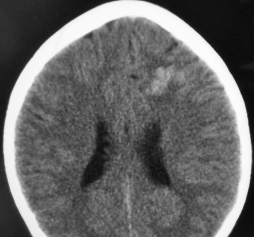
- Grade 1 AVM. a) Cerebral CT showing high density in left frontal lobe.

- Figure 7b
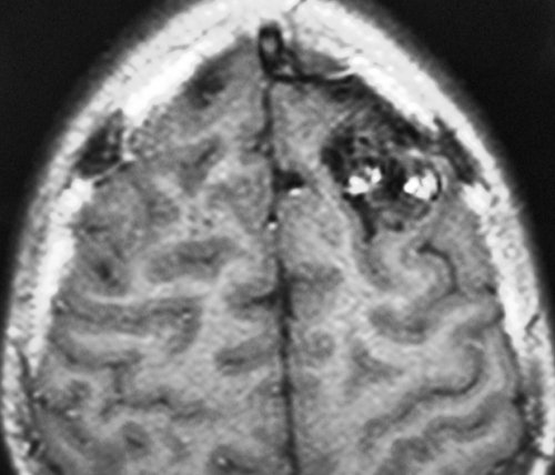
- Grade 1 AVM. b) Axial cerebral T1-weighted MRI showing flow void corresponding to a left frontal AVM.

- Figure 7c
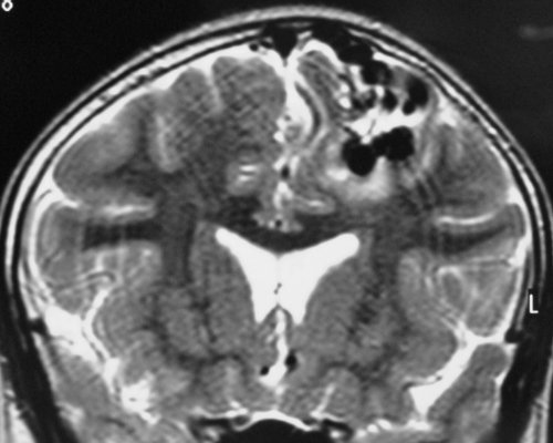
- Grade 1 AVM. c) Coronal cerebral T2-weighted MRI showing flow void corresponding to a left frontal AVM.


- Figure 7d
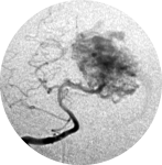
- Grade 1 AVM. d) Angiography in an antero-posterior (AP) projection showing left frontal AVM fed by left anterior cerebral artery (ACA) branches and draining to the superior sagittal sinus.

- Figure 7e
- Grade 1 AVM. e) Postoperative angiography in an AP projection showing complete ressection of the AVM.
- Figure 7f
- Grade 1 AVM. f) Postoperative angiography in an lateral projection showing complete ressection of the AVM.

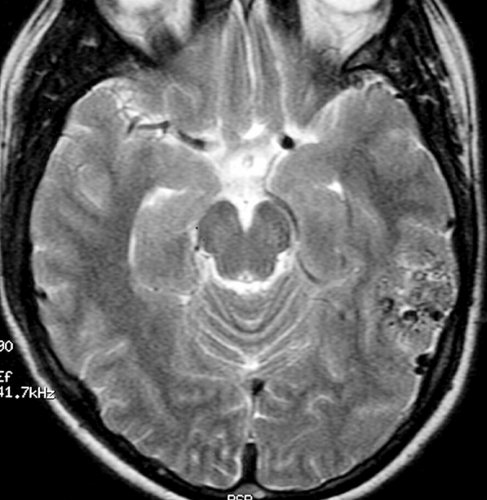
- Figure 8a
- Grade 2 AVM. a) Axial cerebral T2-weighted MRI showing flow void corresponding to a left temporal AVM.

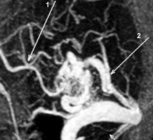
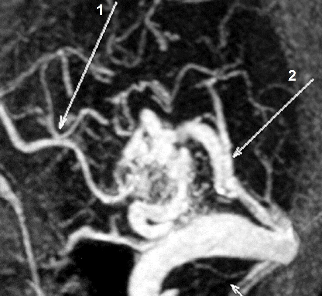
- Figure 8b
- Grade 2 AVM.
b) Computed tomography angiography (CT angiography) showing an AVM fed by a branch of the left posterior cerebral artery (PCA) (arrow 1) and draining to the sigmoid sinus.
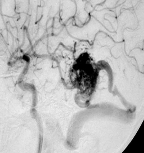
- Figure 8c
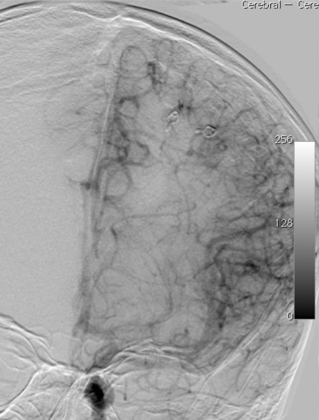
- Grade 2 AVM. c) Angiography in a lateral projection showing an AVM fed by a branch of the left PCA.

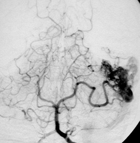
- Figure 8d
- Grade 2 AVM. d) Angiography in an AP projection showing an AVM fed by a branch of the left PCA.

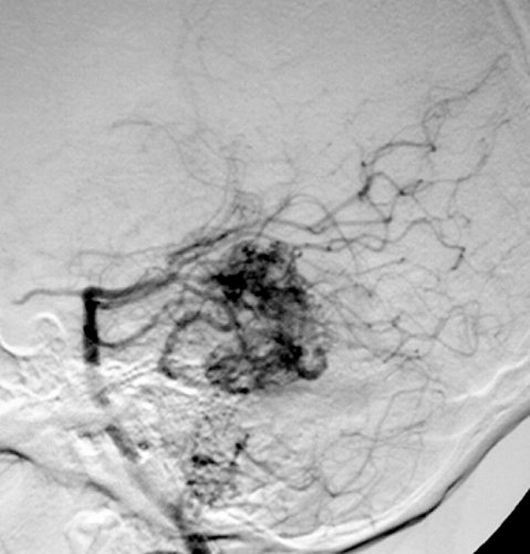
- Figure 8e
- Grade 2 AVM. e) Angiography in a lateral oblique projection showing an AVM fed by a branch of the left PCA and draining to the sigmoid sinus.

- Figure 8f
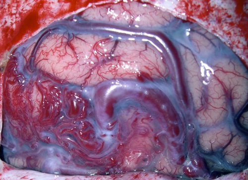
- Grade 2 AVM. f) Intraoperative view of the AVM.


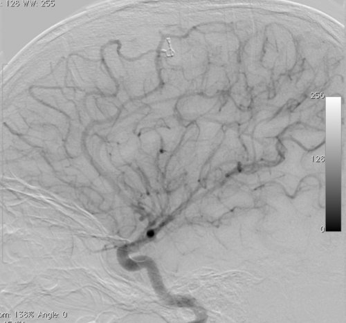
- Figure 8g
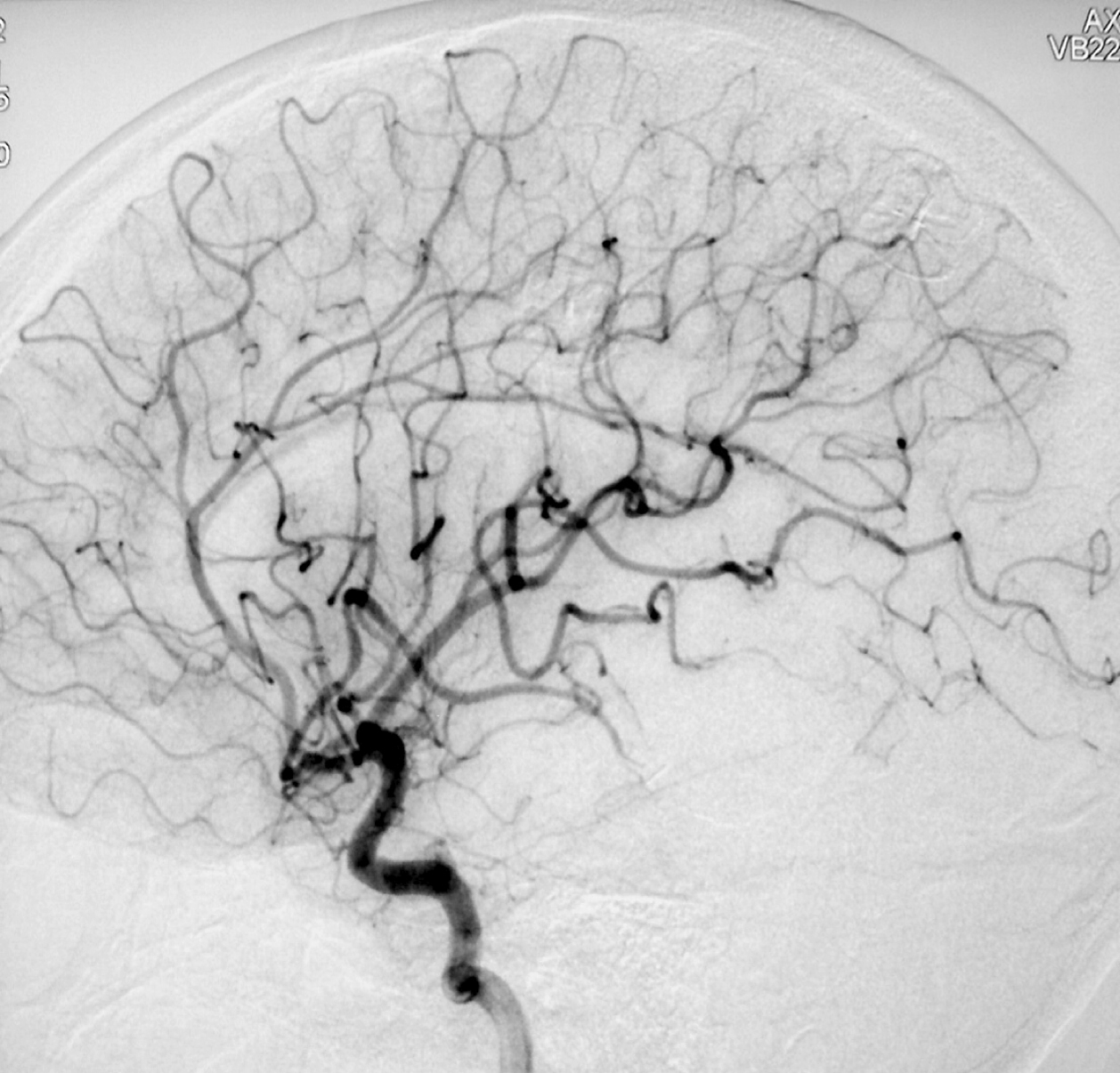
- Grade 2 AVM. g) Postoperative angiography of the left internal carotid artery in a lateral projection showing complete ressection of the AVM.
- Figure 8h
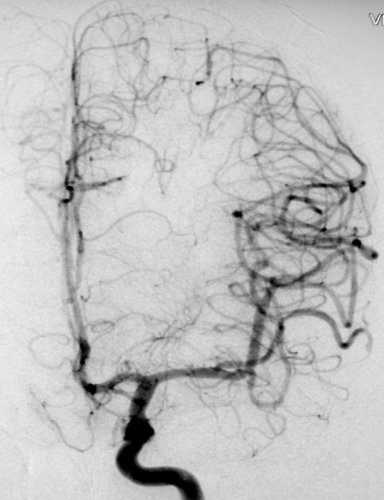
- Grade 2 AVM. h) Postoperative angiography of the left internal carotid artery in an AP projection showing complete ressection of the AVM.

- Figure 8i
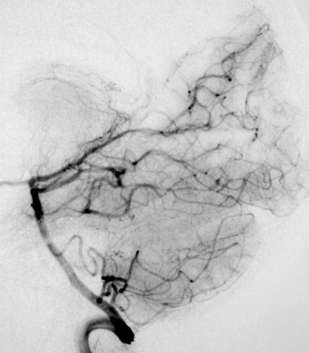
- Grade 2 AVM. i) Postoperative angiography of the left vertebral artery in a lateral projection showing complete ressection of the AVM.
